
Skin Sensitization ISO 10993-10
Overview of Skin sensitization testing
Currently, three animal test methods are used for evaluating the skin sensitization potential of cheMICal substances: two guinea pig methods and one mouse method. Among them, the most commonly used are the Guinea Pig Maximization Test (GPMT) and the Buehler Test.
GPMT is the most sensitive method, the Buehler Test is most suitable for topically applied products, and the Mouse Local Lymph Node Assay (LLNA) is an internationally recognized method for evaluating individual chemical substances. This document outlines the testing principles, sample and animal preparation, test procedures, observations, and evaluation criteria for each method.
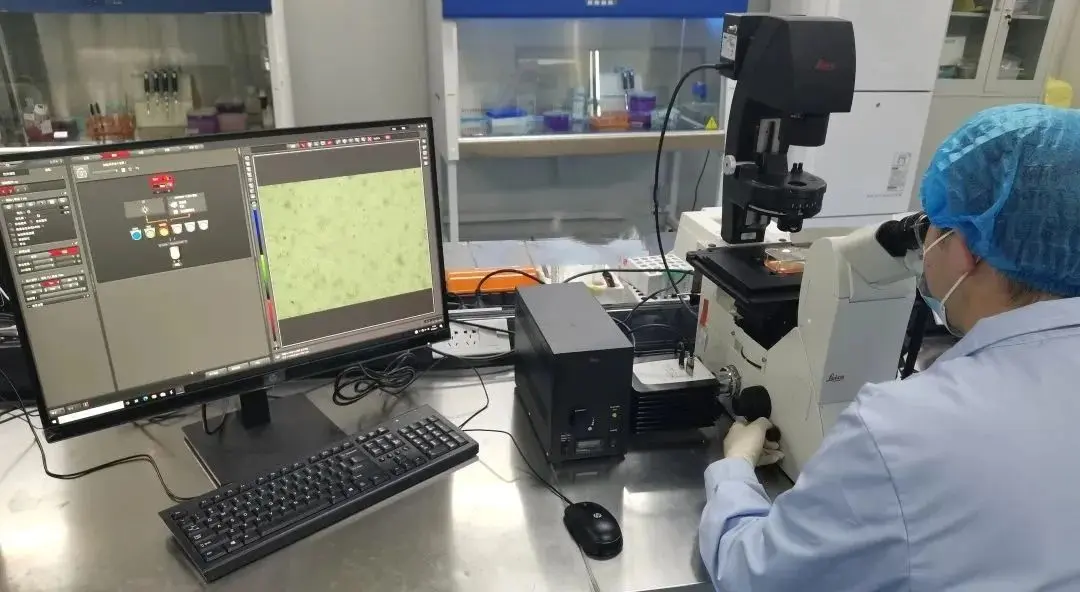
Mouse Local Lymph Node Assay (LLNA)
1. Test Principle
The test sample is applied to the dorsal surface of the mouse's ears, and the degree of lymphocyte proliferation in the lymph nodes (excluding the ear itself) is measuRED. If the proliferation in the test group is three times or more than the control group, the material is considered a sensitizer. Undiluted extracts may be used for medical device testing.
2. Sample and Animal Preparation
Test samples can be in the form of liquids, suspensions, gels, or viscous substances applied to the mouse’s ear. Typically, a concentration series is used; otherwise, the highest concentration is applied. The common extraction vehicle is a 4:1 mixture of acetone and olive oil (AOO). For hydrophilic liquids that do not adhere well to the skin, 0.5% (w/v) carboxymethylcellULose or hydroxyethylcellulose may be added. For water-soluble substances, dimethyl sulfoxide (DMSO) or dimethylformamide (DMF) may be added.
All extraction vehicles or additives must be validated and documented to ensure they do not affect the test results.
The preferred test animals are healthy, non-pregnant, 8–12-week-old female CBA/Ca or CBA/J mice. Other acceptable strains include DBA/2, B6C3F1, and BALB/c, matched within a one-week age range. Animal selection and housing must comply with ISO 10993-2.
3. Test Procedure
For chemical substances, LLNA is typically performed using a dose-response approach. For medical device extracts, a single-dose test without dilution is commonly used. However, for highly toxic extracts, dilution and dose-response testing are recommended due to potential false negatives.
To ensure reproducibility and sensitivity, weak to moderate contact allergens such as mercaptobenzothiazole, hexyl cinnamic aldehyde, and benzocaine may be used. Body weight and clinical observations should be recorded throughout the study.
Each test group should include at least four mice. If only one dose is tested, a minimum of five mice per group is required. Lymph node responses may be measured using individual or pooled samples.
4. Cell Proliferation Assessment
Proliferating cells in the lymph nodes are labeled with radioactive or fluorescent markers. Common radioactive markers include ^3H-methyl thymidine and ^125I-iododeoxyuridine. Fluorescent labeling may involve fluorodeoxyuridine.
At 72 ± 2 hours after the final treatment, mice are weighed and injected intravenously with the marker. At 5 ± 0.75 hours post-injection, the mice are euthanized, and auricular lymph nodes are collected. A single-cell suspension is prepared using a 200 μm steel or nylon mesh. The suspension is washed twice and resuspended in PBS. Cells are precipitated with 5% TCA at 4 ± 2°C for 18 ± 1 hours. After centrifugation, the precipitate is resuspended in 1 mL TCA and transferred to scintillation vials with 10 mL scintillation fluid for ^3H counting or directly to a gamma counter for ^125I counting.
5. Results and Discussion
Radioactivity levels are measured as counts per minute (cpm) per mouse and converted to disintegrations per minute (dpm). The average and standard deviation of cpm or dpm per group are calculated, background values are subtracted, and the Stimulation Index (SI) is determined. An SI ≥ 3 in three or more samples is considered a positive result.
Guinea Pig Maximization Test (GPMT)
1. Sample and Animal Preparation
Samples should be prepared as per Appendix A of the standard and tested at the highest concentration that does not compromise the test. Healthy albino guinea pigs weighing 300–500 g, from a single outbred strain and either male or non-pregnant females, should be used.
Each test group should contain at least 10 guinea pigs and the control group at least 5. If ambiguous or uncertain results occur, testing should be repeated with at least 20 test animals and 10 controls. Animal housing and selection should comply with iso 10993-2.
2. Test Procedure
Preliminary Test
Conducted to determine the appropriate concentration for the main test. Diluted samples are topically applied to the flank (at least 3 animals). After 24 hours, dressings are removed and application sites are graded for erythema and edema. Choose the highest concentration causing mild to moderate erythema for the induction phase and the highest non-irritant concentration for the challenge phase.
Intradermal Induction
Each animal receives 0.1 mL intradermal injections in the scapular region:
Site A: FCA and vehicle in 1:1 volume ratio.
Site B: Undiluted test sample (vehicle for controls).
Site C: Test sample at Site B concentration mixed with FCA in 1:1 ratio (controls receive vehicle + FCA mixture).
Topical Induction
Seven days later, apply \~8 cm² patches soaked with the test sample to the injection sites. If the chosen concentration does not cause irritation, pre-treat the area with 10% SDS for 24 ± 2 h. Dressings are removed after 48 ± 2 h.
Challenge Phase
Fourteen days after topical induction, apply the test and control samples to untreated areas (e.g., upper abdomen) using patches at Site C concentration. Dressings are removed after 24 ± 2 h.
3. Animal Observation
At 24 ± 2 h and 48 ± 2 h post-removal, observe challenge sites under full-spectrum light. Grade erythema and edema responses for each site and time point.
4. Result Evaluation
If the control score <1 and test score ≥1 → sensitizer.
If control score ≥1 and test reaction is stronger → sensitizer.
Suspected responses should be confirmed by re-challenge.
Buehler Test
1. Sample and Animal Preparation
Same as section 3.1 (GPMT).
2. Test Procedure
For all topically applied products, saturate appropriate-sized patches (filter paper or absorbent gauze) with the test material or extract and apply to clipped areas under occlusion for 6 ± 0.5 h.
Preliminary Test
Apply four concentrations to flanks of at least three animals using occlusive dressings. After 6 ± 0.5 h, remove patches and assess erythema and edema at 24 ± 2 h and 48 ± 2 h post-removal. Choose:
Induction: Highest concentration causing no erythema.
Challenge: Highest concentration not causing irritation.
Induction Phase
Apply soaked patches to the upper left back of each animal for 6 ± 0.5 h, three times weekly for three weeks. Controls receive vehicle patches.
Challenge Phase
Fourteen days after the last induction, challenge all animals with test sample on a previously untreated, shaved site. Remove dressings after 6 ± 0.5 h.
3. Animal Observation
Observe and grade test sites at 24 ± 2 h and 48 ± 2 h post-removal under natural or full-spectrum light.
4. Result Evaluation
If the control score <1 and test score ≥1 → sensitizer.
If control score ≥1 and test reaction is stronger → sensitizer.
Suspected responses should be confirmed by re-challenge.
References
iso 10993-10:2010 Biological evaluation of medical devices -- Part 10: Tests for irritation and skin sensitization
Email:hello@jjrlab.com
Write your message here and send it to us
 IEC 62471 Photobiological Safety of Lamps and Lamp
 New European Toy Standard EN 71-1:2026
 EN71 Series Standards Compliance February 13, 2026
 European Toy Safety Standard EN 71-20:2025
 EN 18031 Certification for Connected Devices on Am
 Compliance Guide for Portable Batteries on Amazon
 2026 EU SVHC Candidate List (253 Substances)
 LFGB Certification Cost and Timeline Guide
Leave us a message
24-hour online customer service at any time to respond, so that you worry!
